
Clinical EvidencePRELUDE Study - ATK
Clinical Outcomes
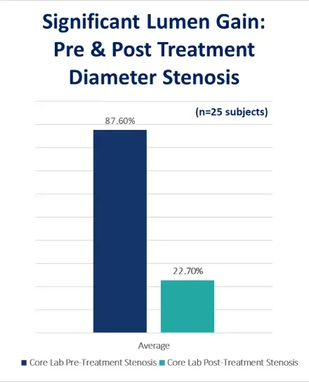
The initial outcomes demonstrate that the Serranator can achieve excellent lumen gain, with minimal dissection and low reintervention rates.
Final Residual Stenosis
Bailout Stent Rate
Freedom from CD-TLR at 6 months
Prospective, core-lab adjudicated
The Serranator® met its safety and efficacy endpoints in a preliminary study for the treatment of femoro-popliteal disease. There was 100% device technical success. The acute results of this feasibility study demonstrate that the Serranator® can safely achieve low residual stenosis. Successful lumen gain was achieved with an average post diameter stenosis of 23%, from a pre-procedure of 88%. In a majority of subjects (68%), the lumen gain was achieved with only 6 ATM of pressure. Serrations were documented by OCT or IVUS in all subjects (n=10) who underwent post Serranator imaging.
(NCT03001700) – https://journals.sagepub.com/doi/pdf/10.1177/1526602818820787
Serration Evidence
As part of the PRELUDE Studies, optical coherence tomography (OCT) and intravascular ultrasound (IVUS) were performed on a sub-set of cases. In all cases, imaging demonstrated serrations regardless of plaque morphology. (n=10/10 subjects)
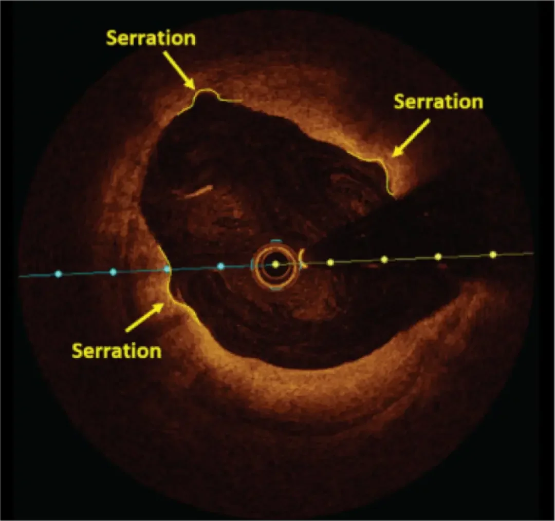
OCT: “Shows clear evidence of serration caused by the Serranator® device.”
– A. Holden, MBChB, FRANZCR, EBIR
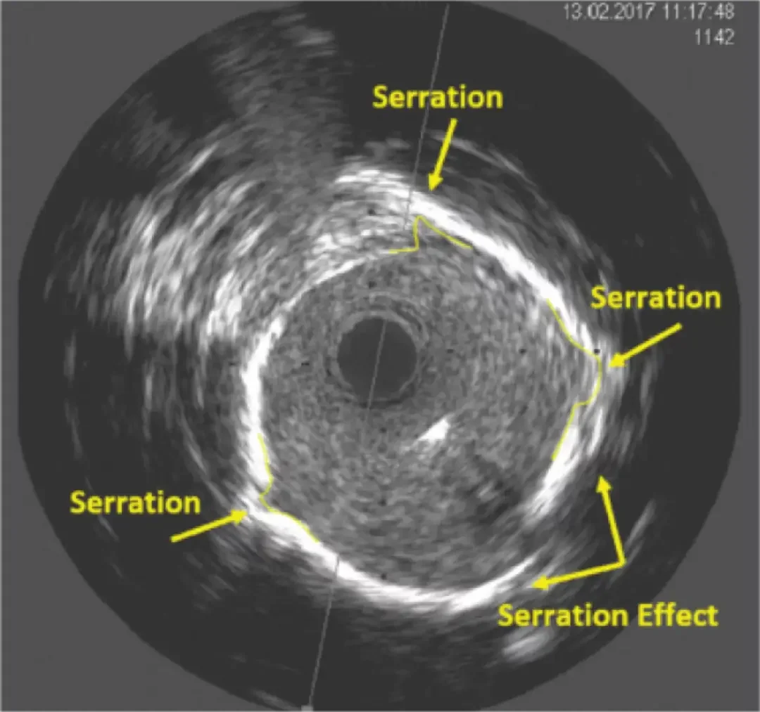
IVUS: “Controlled modification of severe intimal calcification by the Serranator®. Note the controlled acute luminal gain of the impacted calcified intimal layer.”
– J. Mustapha, MD
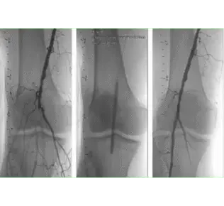
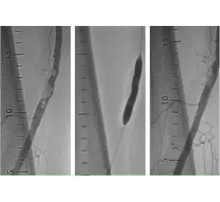
Right Proximal Popliteal with Total Occlusion
RVD: 4.19mm
Lesion length: 54.28mm
Stenosis: 100%
Serranator® size: 5 x 80mm
Inflation pressure: 6 ATM
Post-treatment residual stenosis: 20.23%
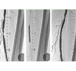
Right Distal SFA with Severe Calcification
RVD: 6.12mm
Lesion length: 30.04mm
Stenosis: 94.59%
Serranator® size: 6 x 40mm
Inflation pressure: 11 ATM
Post-treatment residual stenosis: 24.07%
Left Mid SFA
RVD: 5.15mm
Lesion length: 28.42mm
Stenosis: 77.02%
Serranator® size: 5 x 40mm
Inflation pressure: 6 ATM
Post-treatment residual stenosis: 12.84%
