
Clinical EvidencePRELUDE Study - BTK
Clinical Outcomes
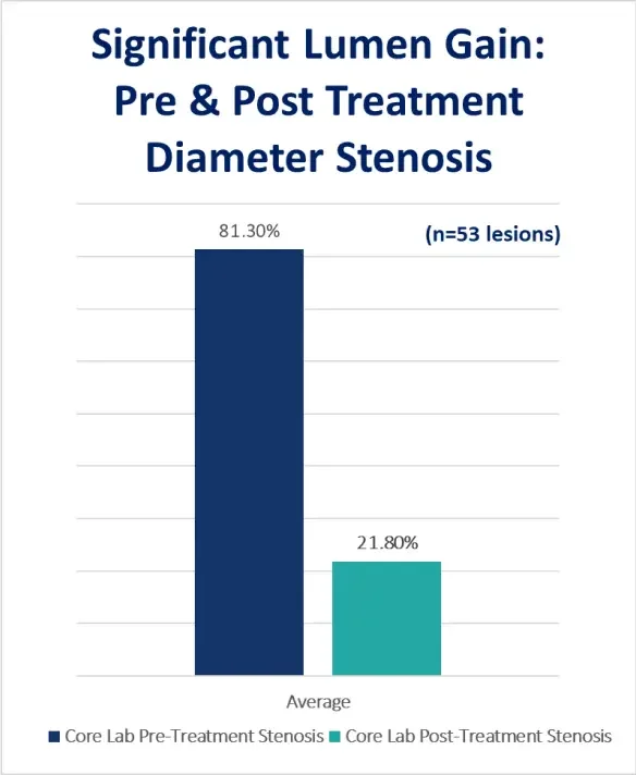
The initial outcomes demonstrate that the Serranator can achieve excellent lumen gain, with minimal dissection and low reintervention rates. (n=53 lesions)
Final Residual Stenosis
Maximum Pressure (mean)
Bailout Stent Rate
Freedom from CD-TLR at 6 months
Additionally, dramatic quality of life and Rutherford classification improvements were demonstrated.Prospective, core-lab adjudicated
The Serranator was found to be safe and effective in treating atherosclerotic disease of the infrapopliteal arteries. The PRELUDE BTK study also showed effective lumen gain with an average pre-procedure diameter stenosis of 81.3%, and an average post treatment diameter stenosis of 21.8%. The lumen gain was achieved using low atmospheric pressure (avg maximum of 6ATM). The mechanism of action was documented by OCT and IVUS. The Serration Angioplasty effect was shown in all 10 lesions imaged. A low re-intervention rate was observed. The Freedom from CD-TLR at 6-months was 97.7%.
(NCT03693963) – https://journals.sagepub.com/doi/10.1177/15266028211059917
Serration Evidence
As part of the PRELUDE Studies, intravascular ultrasound (IVUS) and optical coherence tomography (OCT) were performed on a sub-set of cases. In all cases, imaging demonstrated serrations regardless of plaque morphology. (n=10/10 lesions)
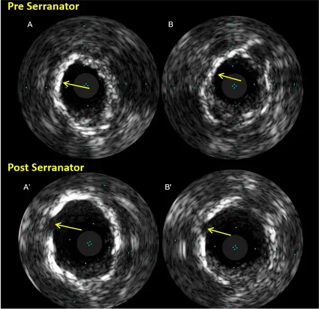
Pre-intervention imaging showed diffuse circumferential superficial calcification (A-B). Post-Serranator imaging showed slits in the superficial calcium enabling enlargement of lumen without any dissection (A’-B’)” – Dr. Akiko Maehara
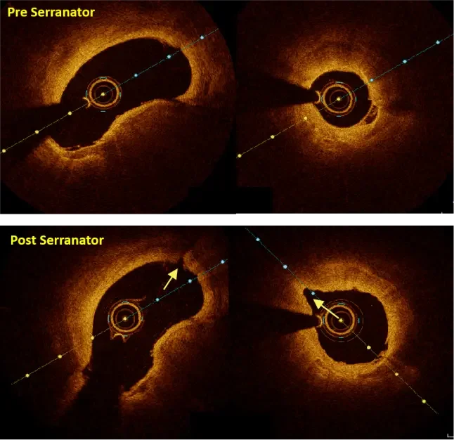
OCT Shows Serrations – Performed by Dr. Andrew Holden
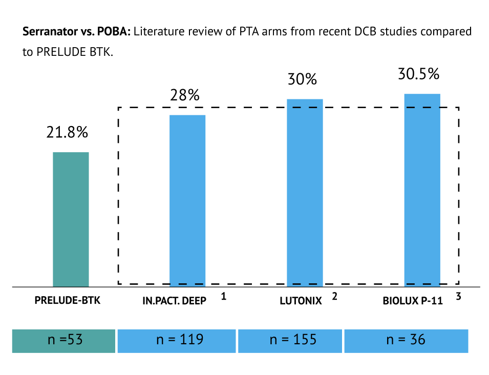
All Data is for POBA trials or POBA control arms from other studies; *Average Represents a Combined Weighted Average including all Studies
1. Zeller, T., Baumgartner, I., Scheinert, D., Brodmann, M., Bosiers, M., Micari, A., . . . Rocha-Singh, K. J. (2014). Drug-Eluting Balloon Versus Standard Balloon Angioplasty for Infrapopliteal Arterial Revascularization in Critical Limb Ischemia. Journal of the American College of Cardiology, 64(15), 1568-1576. doi:10.1016/j.jacc.2014.06.119
2. Mustapha, J., Brodmann, M., Geraghty, P., Saab, F., Settlage, R., & Jaff, M. (2019). Drug-Coated Versus Uncoated Percutaneous Transluminal Angioplasty in Infra-popliteal Arteries: Six-Month Results of the Lutonix BTK Trial. Journal of Vascular Surgery, 70(5), 1718. doi:10.1016/j.jvs.2019.08.011
3. Zeller T, Beschorner U, Pilger E, Bosier M, Deloose k,... Brodmann M. (2015). Paclitaxel-Coated Balloon in Infrapopliteal Arteries: 12-Month Results From the BIOLUX P-II Randomized Trial (BIOTRONIK'S-First in Man study of the Passeo-18 LUX drug releasing PTA Balloon Catheter vs. the uncoated Passeo-18 PTA balloon catheter in subjects requiring revascularization of infrapopliteal arteries).
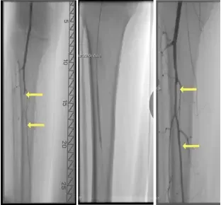
Tibial Peroneal Trunk
Stenosis: 100%
Serranator® size: 3 x 40mm
Maximum ATM: 6
Post residual stenosis: 22%
No dissection
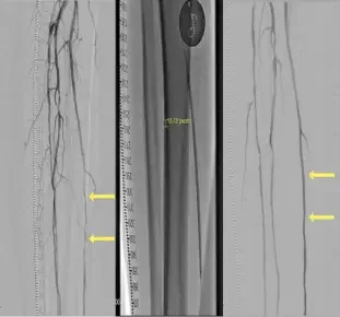
Proximal & Distal Posterior Tibial
Stenosis: 80%
Serranator® size: 3 x 120mm
Maximum ATM: 6
Post residual stenosis: 22%
No dissection
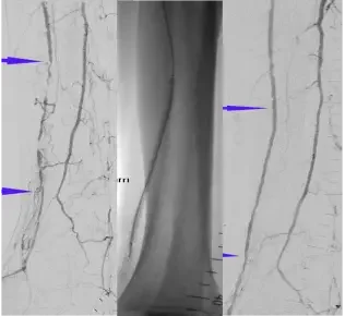
Distal Anterior Tibial
Stenosis: 100%
Serranator® size: 2.5 x 120mm
Maximum ATM: 4
Post residual stenosis: 15%
No dissection
