EVT 2026Achieving Durable Results Above the Knee
A consistent algorithm for complex femoropopliteal disease.
By Michael L. Campbell, MD
Michael L. Campbell, MD | Lakeland Regional Hospital, Lakeland, FL
The femoropopliteal segment is the longest arterial segment in the leg, subject to flexion, torsion, and compression, and it carries a high burden of calcification and long-segment disease. Restoring flow is rarely the whole objective. The harder question is how to restore it in a way that can be maintained and revised when it eventually fails.
Conventional balloon angioplasty applies undirected radial force to the entire vessel wall at once, so the operator has limited control over where and how the artery gives way. Serration Remodeling Therapy (SRT) with the Serranator® PTA Serration Balloon (Cagent Vascular) changes where that force is applied. Serration strips embedded along the balloon create longitudinal micro-fissures within the plaque at low pressure, so the artery opens along defined lines rather than wherever it happens to be weakest. The result is a smooth and uniformly enlarged lumen, and an artery remodeled for whatever treatment comes next.
TREATMENT ALGORITHM
SRT is the constant in my above-the-knee (ATK) algorithm. What changes with lesion morphology is what happens before it, not whether it is used. In fibrotic and mixed disease, I go directly to SRT. In heavy calcification and chronic total occlusions, including previously stented segments, I debulk first with laser atherectomy and then perform SRT across the full treated length. Drug delivery follows in both.
Soft plaque, mixed plaque, and dense calcification all respond to the same controlled mechanism, and in each I finish with a smooth and uniformly expanded lumen at low pressure. Drug-coated balloon (DCB) therapy depends on contact with the vessel wall, and remodeling first gives it a consistent surface to work against.
Case 1
Presentation
A woman in her early 60s with type 1 diabetes, hypertension, coronary artery disease, and prior cerebrovascular accident was referred for a necrotic left lateral foot and heel wound of at least 5 months with associated cellulitis. Pedal pulses were nonpalpable, and she had never undergone a vascular workup. Duplex demonstrated monophasic flow throughout the left leg with no detectable flow in the dorsalis pedis artery (DPA).
Procedural Overview
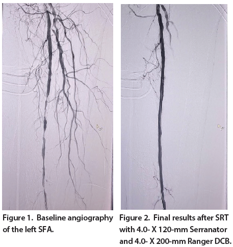
Left lower extremity runoff angiography demonstrated diffuse long-segment disease of the superficial femoral artery (SFA) from the ostium throughout its course (Figure 1). The popliteal artery was patent, and the leg was supplied by single-vessel anterior tibial (AT) artery runoff.
SRT was performed across the diseased SFA segment using a 4.0- X 120-mm Serranator PTA Serration Balloon, followed by angioplasty with a 4.0- X 200-mm Ranger™ DCB (Boston Scientific Corporation) (Figure 2).
Procedural Results
Completion angiography demonstrated markedly improved luminal gain throughout the SFA without significant residual stenosis. No stent was required and no flow-limiting dissection was encountered. Single-vessel AT arterial flow was preserved with good flow through the pedal plantar loop.
A long, diffusely diseased femoral segment was opened gently and remodeled along its entire length, producing the uniform lumen I had aimed for before drug was delivered.
Case 2
Patient Presentation
A woman in her late 70s weighing 38 kg was admitted with a nonhealing left lower extremity wound and progressive cellulitis. Her history included chronic obstructive pulmonary disease on home oxygen, chronic hypoxic respiratory failure, atrial fibrillation, heart failure with preserved ejection fraction, pulmonary hypertension, and treated lung cancer. Revascularization had been deferred during an earlier admission and remained pending when she returned with a worsening wound.
Procedural Overview
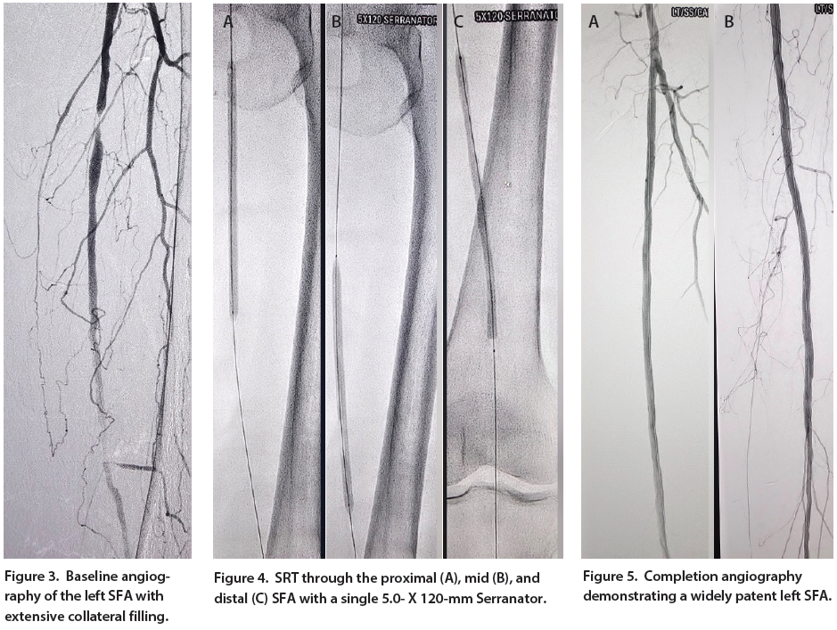
Angiography demonstrated moderate to severe stenosis of both common iliac arteries (CIAs) and multifocal stenotic lesions throughout the left SFA (Figure 3). The popliteal, posterior tibial, and peroneal arteries were patent, with no imaged flow in the DPA.
Inflow was addressed first and covered stenting of both CIAs. SRT was then performed throughout the entire course of the SFA using a single 5.0- X 120-mm Serranator, treated sequentially from proximal to distal (Figure 4).
Procedural Results
Completion angiography demonstrated a widely patent SFA without significant residual stenosis and no flow-limiting dissection (Figure 5). No implant was left in the femoropopliteal segment. The DPA, not imaged at baseline, was intact on completion. Total contrast was 40 mL with an air kerma dose of 117 mGy.
In a patient with minimal physiologic reserve, one Serranator opened the entire diseased SFA at low pressure, restoring a uniform lumen without dissection and leaving nothing behind in a vessel that flexes with every step.
Conclusion
Successful ATK intervention depends on opening the artery in a way it can tolerate and preparing it for the therapy that follows. SRT provides a controlled method of expanding and remodeling the vessel that behaves consistently across the full range of femoropopliteal morphology, achieving predictable expansion while preserving the artery and the options that come with it.