
EVT 2026SRT for EVAR: Achieving Large-Bore Delivery Access for EVAR in Severely Underexpanded Iliac Stents
Dr. Mazin Foteh, MD, FACS, FSVS
Mazin Foteh, MD | Baylor Scott & White The Heart Hospital, Plano, TX
Case Presentation
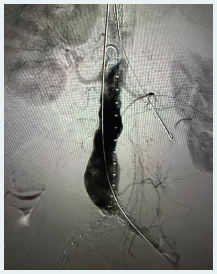
Figure 1. Pre-procedure angiogram.
A woman in her early 80s with known aortoiliac occlusive disease presented for repair of an abdominal aortic aneurysm measuring 5.4 cm in maximum diameter. She had undergone prior bilateral iliac stenting. Imaging demonstrated severe in-stent restenosis bilaterally with chronic total occlusion of the left external iliac artery, and the existing stents were markedly underexpanded (Figure 1). The combination of dense neointimal hyperplasia and residual underexpansion produced an effective luminal diameter well below that required to advance the endograft delivery system. Given her age, iliac conduit creation or open repair carried substantial morbidity, and a totally endovascular solution was preferred.
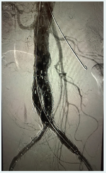
Figure 2. Post-procedure angiogram.
Procedural Overview
Bilateral femoral access was obtained. Initial attempts to advance a large-caliber sheath failed on both sides, with the system arresting at the underexpanded stented segments. Sheath sizing was staged to what the vessels would accept: 16-F on the right and 14-F on the left.
The left external iliac occlusion was crossed intraluminally. Serration Remodeling Therapy (SRT) was performed bilaterally using 8.0- X 40-mm Serranator® PTA Serration Balloon Catheters (Cagent Vascular), one per iliac, with prolonged low-pressure inflations.
Both systems subsequently accepted the delivery sheaths, and a 36- X 80-mm Treo® Abdominal Stent-Graft System (Terumo Aortic) was deployed to exclude the aneurysm. On the postimplant CT scan, the iliac limbs demonstrated excellent expansion with no evidence of recurrent stenosis (Figure 2).
WHY SRT?
The prior iliac stents were both densely restenosed and significantly underexpanded, leaving a lumen that would not permit safe passage of a large-bore sheath. SRT was selected for its serration mechanism: the strips concentrate point force along the neointima, disrupting hyperplastic tissue at low inflation pressure rather than stretching it.
The disease state itself narrowed the options. Conventional angioplasty at the inflation pressures required to move a stent that had never fully expanded carries real risk of vessel injury or rupture, and dense neointima tends to recoil once the balloon comes down. Intravascular lithotripsy is designed to modify calcium and does little to hyperplastic tissue. Atherectomy within a stented segment introduces the risk of damage to the stent frame or device entrapment. SRT was the one modality that addressed both components of this lesion, the neointima and the stent geometry, in a single treatment at low inflation pressure, which made it the appropriate therapy for this anatomy.
That same point force allowed the underexpanded stents to be expanded further. Together, this created a durable delivery corridor and enabled a successful endovascular repair in a patient for whom iliac conduit or open conversion would have carried substantial morbidity.
“SRT was selected for its serration mechanism: the strips concentrate point force along the neointima, disrupting hyperplastic tissue at low inflation pressure rather than stretching it.”