
EVT 2026Pedal Disease: Can Vessel Remodeling Improve Distal Revascularization?
By Melissa Keller, MD, PhD
Melissa Keller, MD | UC Davis Health, Sacramento, CA
Case Presentation
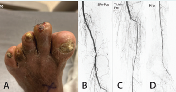
A male diabetic patient in his mid-60s with a history of prior lower extremity intervention presented with a new, nonhealing toe wound (Figure 1A). Although a previous toe wound and amputation had healed following revascularization, the posterior tibial (PT) artery had subsequently reoccluded. Angiography demonstrated significant tibial and inframalleolar disease with essentially single-vessel runoff through the anterior tibial (AT) artery (Figure 1B-1D). The goal was to re-establish PT and plantar flow to improve perfusion to the foot and address the stenosis in the AT artery.
Procedural Overview
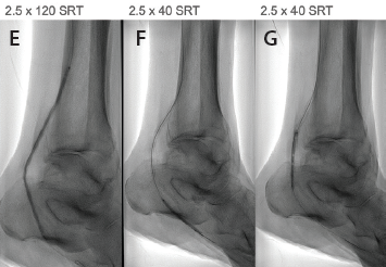
Using a 6-F, 65-cm Destination® sheath and a 0.014-inch Glidewire® Advantage (both Terumo Interventional Systems), the PT was successfully recanalized into the lateral plantar artery. After initial plain old balloon angioplasty (POBA), Serration Remodeling Therapy (SRT) with a 2.5- X 120-mm Serranator® PTA Serration Balloon (Cagent Vascular) was utilized through the plantar and PT arteries, with additional SRT treatment using a 2.5- X 40-mm Serranator of a resistant focal segment to optimize expansion (Figure 1E-1H).
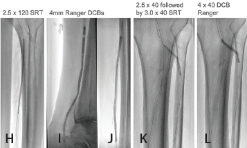
No recoil or waist was observed. In the PT, drug-coated balloon (DCB) therapy using overlapping 4.0-mm Ranger™ DCB (Boston Scientific Corporation) was then used subnominal more distally (Figure 1I and 1J). The AT artery was then treated using a similar algorithm with a 3.0- X 40-mm Serranator as vessel prep, followed by a 4- X 40-mm Ranger DCB (Figure 1K and 1L).
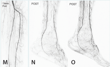
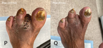
Completion angiography demonstrated improved AT flow, restored PT flow, and improved filling into the foot (Figure 1M-1O). At follow-up toe-brachial index doubled at 2.5 (Figure 1P) and 5 months (Figure 1Q), the wound progressively healed and ultimately remains healed.
Why SRT?
I use SRT across a range of peripheral interventions and have found it particularly valuable when treating tibial and pedal disease. These are some of the smallest and most challenging vessels we encounter, specifically those patients with chronic limb-threatening ischemia, often with a comorbidity of diabetes contributing with diffuse disease extending deep into the inframalleolar region. In this setting, improvement in blood flow is critical for wound healing and limb preservation, and there is very little margin for an unpredictable and ineffective angioplasty result.
What I appreciate about SRT in these vessels is using its controlled vessel remodeling to achieve meaningful luminal expansion without the usual recoil or residual waist. I routinely see a favorable angiographic result with improved flow while maintaining the integrity of the native vessel. That combination is important anywhere I use SRT, but it becomes especially valuable below the ankle, where every millimeter of lumen—and every additional small vessel branch of recruitment for perfusion—can matter.